ATI TEAS 6 - Science (Human Anatomy and Physiology)
Hierarchy of Structures
Lowest Hierarchy level is at Organelles within a cell. They obtain
... [Show More] energy from food and reproduction.
-Cells with the same function are collected into larger groups called Tissues.
-Tissues are collected into Organs, carry out single task, like oxygenated blood (lungs), or filter out waste (kidneys).
-Organs work together in systems that perform coordinated large-scale functions,
like nourishing the body (digestive) or protecting the body from attacks (immune).
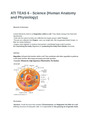
Cell Parts
Organelles: Cell parts that function within a cell. They coordinate with other organelles to performs a cell's basic function, like energy processing and waste excretion.
~Examples: Ribosomes, Golgi Apparatus, Mitochondria, The Nucleus.
The Nucleus
-Nucleus: Small structure that contains Chromosomes and Regulates the DNA of a cell. Defining structure of eukaryotic cells. It is responsible for the passing on of genetic traits between generations.
-Contains: nuclear envelope, nucleoplasm, a nucleolus, nuclear pores, chromatin, and ribosomes.
Chromosomes
Highly condensed, threadlike rods of DNA. DNA is genetic material that stores information about the plant or animal.
Chromatin
Consists of the DNA and Proteins that make up chromosomes.
Nucleolus
Structure contained within the nucleus, consists of proteins. Small, Round, and
does not have a membrane. Involved in protein synthesis, and synthesizes and stores RNA.
Nuclear Envelope
Encloses the nucleus. Consists of inner and outer membranes made of lipids.
Nuclear Pores
Involved in exchange of material between nucleus and the cytoplasm.
Nucleoplasm
Liquid within the membrane and is similar to cytoplasm.
Cell Membrane
"Plasma Membrane"
-Made of Lipids and Proteins
-Isolates the cell from its external environment while still enabling the cellar to communicate with the outside environment.
-Consists: Phospholipid bilayer with the hydrophilic ends of the outer layer facing external environment.
~Cholesterol: Adds stiffness and flexibility
~Glycolipids: Help cell to recognize other cells of the organisms.
~Proteins: Help give cells shape
~Special Proteins: Helps cell communicate with external environment.
~Other Proteins: Transport molecules across membrane
Selective Permeability
With regard to size, charge, and solubility.
-Size: Membrane allows small molecules to diffuse through it. Oxygen and Water molecules are small and can pass through the cells membrane.
-Charge: Ions on a cells surface either attracts or repels ions. Ions with like charges are repelled, and ions with opposite charges are attracted to the surface.
-Solubility: Molecules that are soluble in phospholipids can usually pass through the membrane. Many are not able to diffuse the membrane, and if anything they'll have to be moved through by active transport and vesicles
Cell Structures
Inside the cell. Contain: Ribosomes, Golgi Apparatus, Vacuoles, Vesicles, Cytoskeleton, Microtubules, Cytosol, Cytoplasm, Cell Membrane, Endoplasmic Reticulum, Mitochondria
Ribosomes
Involved in synthesizing proteins from amino acids.
-Make up about a quarter of a cell.
-Some are embedded in the Rough Endoplasmic Reticulum (Rough ER)
Golgi Apparatus
Involved in synthesizing materials like proteins that are transported out of the cell.
~Modifies and Packages proteins secreted from the cell.
-Located near the nucleus and has layers of membranes.
Vacuoles
Sacs used for storage, digestion, and waste removal.
-Plant: Has one large vacuole
-Animal: Has small, sometimes numerous vacuoles.
Vesicles
Small organelle within a cell, has a membrane.
-Functions: Moving materials within a cell.
Cytoskeleton
Consist of microtubules that help shape and support the cell.
Microtubules
Part of cytoskeleton.
-Help support the cell.
-Made of proteins
Cytosol
Liquid materials in the cell. Mostly water, also contains floating molecules.
Cytoplasm
Refers to Cytosol and the substructures (organelles) found within the plasma membrane, but not within the nucleus.
Cell Membrane
Acting as a barrier. Helps keep cytoplasm in and substances located outside the cell out.
-Helps determine what is allowed to exit and enter.
Endoplasmic Reticulum
Two Types:
-Rough ER: Has ribosomes on surface.
~Functions: Manufacture lysosomal enzymes, Manufacture of secreted proteins. (Protein production, protein folding, quality control, and despatch)
-Smooth ER: Has no ribosomes.
~Functions: Manufacture Lipids (fat), Metabolism, Steroid Hormone production (adrenal cortex and endocrine glands), Helps liver detox.
-Tubular Network that comprises the transport system of a cell. It is fused to the nuclear membrane and extendsthrough cytoplasm to the cell membrane.
Mitochondria
Vary in terms of size and quantity. Has various functions.
-Functions: Production of Cell Energy (ATP) (Main function), Cell Signaling (Communications are carried out), Cell Differentiation (Cell transforms into a cell with more specialized purpose), Cell Cycle and Growth Regulation (Growth and Death, Reproduction).
-Inner and Outer membrane:
~Inner: Encloses the matrix. Contains mtDNA and ribosomes.
~Between the 2 Membranes: Cristae (Folds). Chemical reactions occur here that release energy, Control Water Levels in cells, and Recycle and Create Proteins and Fats.
-Aerobic Respiration: Occurs in Mitochondria.
Animal Cell Structure
Contains: Centrosomes, Centriole, Lysosome, Cilia, Flagella Centrosome
Pair of centrioles located at right angles to each other and surrounded by protein.
-Involved in Mitosis and Cell Cycle
Centriole
Cylinder-shaped structures near the nucleus.
-Involved in Cellular Division
-Each cylinder consist of 9 Groups of 3 Microtubules. Occurs in pairs.
Lysosome
-Functions: Digest proteins, lipids, and carbohydrates. Also transports undigested substances to the membrane do they can be removed. Shape depends on material being transported.
Cilia
Appendages extending from the surface of the cell.
-Moves the cell and results in fluid being moved by the cell. Flagella
Tail-like structures on cell that use whip-likemovements to help move the cell. Longer than Cilia. Only has one or a few flagella.
Cell Cycle
The process by which a cell reproduces which involves cell growth, duplication of genetic material, and cell division.
-Complex organisms: Use the cell cycle to replacecells as they lose their functionality and wear out.
-In Animals: Cell Cycle can take 24 hours.
-Human Skin Cells: Constantly reproducing.
-2 Ways for Cell Reproduction: Mitosis and Meiosis
Cell differentiation
Determines the different cell types
-When less-specialized cell becomes a more-specialized cell. Process is controlled by genes of each cell among a group of cells known as a zygote.
-Cell builds certain proteins and other pieces that set it apart as a specific type of cell.
~Example: Gastrulation (early phase in embryonic development in animals) Mitosis
Events that occur: Interphase, Prophase, Metaphase, Anaphase, Telophase, and Cytokinesis.
Interphase
Cell prepares for division by replicating its genetic and cytoplasmic material.
-Further divided into G1, S, G2 (Meiosis) Prophase
-Chromatin thickens into chromosomes and the nuclear membrane begins to disintegrate.
-Pairs of Centrioles move to opposite sides and spindle fibers begins to form.
-Mitotic Spindle moves chromosomes around wishing the cell.
Metaphase
Spindle moves to the center of the cell and chromosome pairs align along the center of the spindle structure.
Anaphase
Pair of chromosomes, sisters, begin to pull apart and may bend. When they separate, they are called daughters. Grooves then appear in cell membrane.
Telophase
Spindle disintegrates, nuclear membranes reform, and the chromosome revert to chromatin.
-Animals Cells: Membrane is pinched
-Plant Cells: New cell wall begins to form
Cytokinesis
Physical splitting of the cell into two cells
- Some believe it occurs following telophase, others say it occurs from anaphase, as the cell begins to furrow, through telophase, when cell actually splits into two.
Meiosis
Same phased as Mitosis, except it happens twice and differentevent occur during some phases.
-First Phase: Interphase(1), Prophase(1), Metaphase(1), Anaphase(1), Telophase(1), and Cytokinesis(1)
-Second Phase: Prophase(2), Metaphase(2), Anaphase(2), Telophase(2), and Cytokinesis(2).
Interphase(1) Divided into 3 Parts:
-G1 Phase: Cell synthesizes proteins, including the enzymes and structural proteins it will need for growth. In G1, each of the chromosomes consists of a single linear molecule of DNA.
-S Phase: The genetic material is replicated; each of the cell's chromosomes duplicates to become two identical sister chromatids attached at a centromere.
-G2 Phase: DNA Replication Prophase(1)
Longest Phase
-Chromosomes cross over, Genetic material is exchanged, and te trades of four chromatids are formed. Nuclear membrane dissolves/breaks down.
Metaphase(1)
Pair of homologous chromosomes move along the metaphase plate. Anaphase(1)
Microtubules shorten, and homologous pairs of chromatids are separated and travel to different poles.
Telophase(1) and Cytokinesis(1)
Pairs arrives at poles and cell is pinched apart, separating into two cells. Prophase(2)
Disappearance of the nucleoli and the nuclear envelope again as well as the shortening and thickening of the chromatids. Centrosomes move to the polar regions and arrange spindle fibers for the second meiotic division.
Metaphase (2)
Centromeres contain two kinetochores (pulls the chromosomes to the poles) that attach to spindle fibers from the centrosomes at opposite poles.
Anaphase (2)
Remaining centromeric cohesion is cleaved allowing the sister chromatids to segregate. The sister chromatids by convention are now called sister chromosomes as they move toward opposing poles.
Telophase (2) and Cytokinesis(2)
Marked by decompensation and lengthening of the chromosomes and the disassembly of the spindle. Nuclear envelopes reform and cleavage or cell plate formation eventually produces a total of four daughter cells, each with a haploid set of chromosomes.
Tissues
Groups of cells that work together to perform a specific function
-Grouped into 4 broad categories: Muscle (Body Movement), Nerve (Brain, Spinal Cord, and Nerves), Epithelial (Layers of Skin/Membranes), and Connective Tissue (Bone tissue, Cartilage, Tendons, Ligaments, Fat, Blood, and Lymph).
~Includes: Epithelial, Connecting, Cartilage, Blood, Bone, Muscle, and Nervous. Epithelial Tissue
Cells are joined together tightly
-Example: Skin Tissue
Connective Tissue
May be dense, loose, or fatty.
-It protects and binds body parts.
Cartilage Tissue
Cushions and provides structural support for body parts.
- Jelly-Like base and is fibrous
Blood
Transports Oxygen to cells and Removes wastes.
-Carries hormones and Defends against disease. Bone Tissue
Hard tissue that supports and protects softer tissues and organs.
-Marrow produces RBC
-Connective Tissue
Muscle Tissue
Helps support and move the body.
-3 Types:
~Smooth: Provides tension in the blood vessels, control pupil dilation, and aid in peristalsis.
~Cardiac: Only found in the heart
~Skeletal: Includes the muscles commonly called biceps, triceps, hamstrings, and quadriceps.
Nervous Tissue
Neurons form a network through the body that control responses to change in the external and internal environment. Some send signals to muscles and glands to trigger responses.
-Located in brain, spinal cord, and nerves
3 Primary Body Planes
-Transverse Plane (Horizontal): Divides the patient's body into upper and lower halves.
-Sagittal Plane (Vertical): Divides the body, or any body part, into right and left sections. Runs parallel to the midline of the body.
-Coronal Plane (Vertical/Frontal): Divides the body, or any body part, into front and back. Runs through the body at right angles.
Terms of Direction
-Medial: Towards the mid-line, Middle, Away from the side.
~Example: The little finger it medial to the thumb.
-Lateral: Toward the side, Away from the mid-line.
~Example: Anatomical position, Thumb is lateral to little finger.
-Proximal: Structures closer to the center of the body.
~Example: Hip is proximal to the knee.
-Distal: Structures further away from center of the body.
~Example: Knee is distal to the hip.
-Anterior: Structures in front.
-Posterior: Structures behind.
-Cephalad/Cephalic: Adverbs meaning towards the head.
~Example: Cranial is the adjective, meaning The Skull.
-Caudad: Adverb meaning towards the tail/posterior.
~Example: Caudal is the adjective, meaning The Hindquarters.
-Superior: Above, or closer to the head.
-Inferior: Below, or closer to the feet.
Organs
Group of tissues that work together to perform specific functions. Organ Systems
Group of organs that work together to perform specific functions.
-Includes: Respiratory, Cardiovascular, Gastrointestinal, Nervous, Muscular, Integumentary, Reproductive, Endocrine, Renal/Urinary, Immune, and Skeletal. Respiratory System Structures
Upper: nose, nasal cavity, mouth, pharynx larynx
Lower: trachea, lungs, and bronchial tree (bronchi, bronchial network)
Airway
- Lined with cilia to remove microbes and debris
-Lungs:
Bronchial Tree -> lungs -> terminate into alveoli (air sacs) -> gas exchange with blood capillaries
Walls of Alveoli allow for the exchange of gases* with the blood capillaries that surround them.
Right lung - 3 Lobes Left lung - 2 Lobes
-Surrounded by Pleural Membrane (reduce friction)
-Muscles:
Diaphragm: separates thoracic/abdominal cavities
Intercostal: between ribs
Respiratory Functions
-Supplies body with oxygen and Removes carbon dioxide (occurs in alveoli)
-Filters Air: passes through nasal passages -> lungs
-Speech: Air -> throat -> through larynx, causing vibrations and producing sound before heading to trachea
-Cough: Particles -> nasal passages/airways -> expelled from body
-Smell: Chemoreceptors (nasal cavity) respond to airborne chemicals Hyperventilation
Increase blood pH during Acidosis (low pH)
Slow breathing during Alkalosis (high pH) -Lowers blood pH Breathing Process
-Diaphragm/Intercostal muscles contracts to expand lungs
-Inspiration (Inhalation): Diaphragm contracts and moves down increasing the chest cavity
-Expiration (exhalation): Intercostal muscles contractand ribs expand, increasing size of chest cavity
~Volume of chest cavity increases, then the pressure inside chest cavity decreases
~When relaxed: Size of cavity decreases forcing air out.
-Controlled by Medulla Oblongata
~Monitors carbon dioxide in blood, signals the breathing rate to increase when levels are too high.
Respiratory Problems
High Altitude: Decrease lung function due to low oxygen levels.
*People who live in high altitude, evolve over time to have larger lungs.
Chemicals, Pollen, Smoke: Damaged cilia causing Emphysema, Allergies, or Inflammation.
Pathogens: Influenza (corona virus), Tuberculosis (mycobacterium), and Pneumonia (walking - mycoplasma infection)
*Mycosis -> Fungus
Cystic Fibrosis (gene mutation), Asthma, Lung Surfactant Insufficiency: Impedes lung action.
Ventilation
Process of aerating the lungs Respiratory Directions
Air -> Trachea -> Bronchi -> Lungs -> Alveoli Alveoli
-Aqueous Surfactant: The median for gas exchange and keeps lungs from collapsing on itself due to surface tension
Pathway of Oxygen/Carbon Dioxide in Lungs
How Respiratory System effects Circulatory System
-Lungs are perfumed by blood vessels from the heart to bring deoxygenated blood rich in carbon dioxide to the lungs, where oxygen is added and carbon dioxide is removed to return oxygenated blood to the heart for circulation to the rest of the of the body
-Diffusion: Passive transport mechanism. Rate of Diffusion is directly proportional to the surface area involved and the concentration gradient, and is inversely proportional to the distance between the two. solutions.
-Oxygen in the lungs moves into the blood, and carbon dioxide in the blood moves to the lungs. Lungs then exhale carbon dioxide back out of body.
Tidal Volume
Amount of air breathed in and out of lungs
Residual Capacity
-Small amount of stale air.
-Remains trapped in alveoli after expiration and mixes with the fresh air brought in by inspiration.
Circulatory System
Internal transport of substances to and from the cells.
-3 Parts: Blood, Blood Vessels, and Heart.
-Open or Closed.
Blood
Human has 5 quarts of blood.
Plasma: Half blood volume. Mostly water, serves as solvent.
-Contains: plasma proteins, ions, glucose, amino acids, hormones, and dissolved gases.
RBC (red): Transports oxygen to cells. Form in bone marrow. Live for 2 Months, constantly replaced.
WBC (white): Defends body against infection and removes waste.
~Lymphocytes, neutrophil, monocytes, eosinophil, and basophil.
Platelets: Fragments of stem cells.
~Function: Blood Clotting
Heart Chambers
4 Chambers: 2 Ventricles, 2 Atriums -Halves separated by AV Valve (located between ventricle and artery leading away from the heart).
Types of Circulation
Coronary: Flow of blood to the heart tissue. Blood enters the coronary arteries, which branch off the aorta, supplying major arteries, which enter the heart with oxygenated blood.
Deoxygenated blood returns to the right atrium through the cardiac veins which empty into the coronary sinus.
Pulmonary: Flow of blood between the heart and the lungs. Deoxygenated blood flows from the right ventricle to the lungs through pulmonary arteries. Oxygenated blood flows back to the left atrium through the pulmonary veins.
Systemic: Flow of blood to the entire body with the exception of coronary and pulmonary. Blood exits the left ventricle through the aorta, which branches into the carotid arteries, subclavian arteries, common iliac arteries, and the renal artery. Blood returns to the heart through the jugular veins, subclavian veins, common iliac veins, and renal veins, which empty into the superior and inferior vena cavae.
-Portal circulation: Included in Systemic. Flow of blood from the digestive system to the liver and then to the heart and renal circulation, which is the flow of blood between the heart and kidneys.
Blood Pressure
Fluid pressure generated by the cardiac cycle.
Arterial: Functions by transporting oxygen-poor blood into the lungs and oxygen-rich blood to the body tissues.
-Arteries branch into smaller arterioles which contract and expand based on signals from the body.
-Arterioles are where adjustments are made in blood delivery to specific areas based on complex communication from body systems.
Capillary Beds: Diffusion sites for exchanges between blood and interstitial fluid. Capillary: Has thinnest wall of any vein, consisting of single cell endothelial cells. Merge into venues which in turn merge with larger diameter tubules called veins.
-Veins transport blood from body tissues back to the hearts. Thin and contain smooth muscle and function as blood volume reserves.
-Valves inside the veins facilitate this transport. Lymphatic System
-Function: To return excess tissue fluid to the bloodstream.
-Consists of transport vessels and lymphoid organs.
Lymph Vascular System: Consists of lymph capillaries, lymph vessels, and lymph ducts.
-Function: Return excess fluid to blood, Return of protein from capillaries, Transport of fats from the digestive tract, Disposal of debris and cellular waste.
Lymphoid Organs
-Consist of lymph nodes, spleen, appendix, adenoids, thymus, tonsils, and small patches of tissue in the small intestines.
Lymph Nodes: Located at intervals through the lymph vessel system. Contains lymphocytes and plasma cells.
Spleen: Filters blood, stores of RBC and macrophages.
Thymus: Secrets hormones. Major site of lymphocyte maturation.
Spleen
-Upper left of the abdomen, behind the stomach and below diaphragm.
-Lymphoid tissue
-Blood vessels are connected to the spleen by splenic sinuses.
-Function: Filter unwanted materials from the blood (including old RBC) and to help fight infections.
-Up to 10% of the population has one or more accessory spleens that tend to form at the hilum of the original spleen.
Peritoneal Ligaments that Support the Spleen
-Gastrolienal: Connects the stomach to the spleen
-Lienorenal: Connects the kidney to the spleen
-Middle Section of the Phenicol ligament: Connects the left colic flex use to the thoracic diaphragm
Heart Functions
-Atrial Contraction: Fills ventricles and then ventricular contractions empty them, forcing circulation "cardiac cycle"
-Cardiac Muscles attach to each other and signals for contraction spreading rapidly.
-Complex Electrical System: Controls the heartbeat
-Cardiac Muscle Cells: Produce and conduct electrical signals. Capillaries
Drain interstitial fluid that fills the spaces between cells
-Filters it through a system of lymph nodes that are enriched in lymphocytes and provide surveillance by immune system.
Lymph
-Essentially plasma with RBCs removed [Show Less]