The heart is the core of the cardiovascular system. This
double pump beats automatically, night and day, to
keep blood circulating around the body. A
... [Show More] heart “beat”
is a sudden tightening of the muscle in the walls of the
heart. This squeezes blood out of the heart chambers
and into the blood vessels. A specialized type of heart
muscle called myocardium gives the heart its special
pumping ability.
Blood for the heart
The heart muscle needs a
generous supply of oxygen to
keep it working efficiently.
This oxygen is delivered by
the two coronary arteries,
which cover the surface of
the heart with a network that
looks rather like a crown. The
term “coronary” comes from
the Latin word “coronarius,”
meaning “belonging to a
crown or wreath.” Structures
and events involving the
heart often make use of the
word “coronary.”
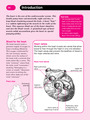
Heart from above
Heart valves
Working within the heart muscle are valves that allow
blood to flow through the heart in only one direction.
The heart valves also prevent the backflow of blood in
the major arteries and veins.
Coronary
arteries
●
●
Upper left
pulmonary vein ●
Right pulmonary
veins
●
●
Aortic valve at
beginning of aorta
●
Pulmonary valve
at beginning of
pulmonary artery
●
Superior
vena cava
●
SECTION 2: HEART 29
© DIAGRAM
Heart facts
• The heart beats some two
and a half billion times
during a 70-year lifetime.
• About five percent of all
the blood pumped by the
heart goes to its own
muscle tissue.
• The heart pumps roughly
3,000 gallons (11,356 l) of
blood in a day.
Arteries and veins of the heart
The largest arteries and veins of the heart
form the beginning of the pulmonary
(lung) and systemic (body) circulations.
Two large veins called venae cavae drain
oxygen-poor blood from the body to the
heart. The pulmonary arteries carry this
blood from the heart to the lungs. The
pulmonary veins carry oxygen-rich blood
from the lungs back to the heart. The
aorta then carries this oxygen-rich blood
to the body.
Heart from behind
Left pulmonary veins
●
●
Left pulmonary artery ●
Inferior vena cava ●
Right pulmonary artery ●
Superior vena cava ●
Left common carotid artery
●
Innominate (brachiocephalic) artery ●
Right pulmonary veins
●
Left subclavian artery ●
Aortic arch
●
●
30 The heart and thorax
Heart beats
To listen to the heart
beating, doctors and
nurses position a
stethoscope between the
fifth and sixth ribs on a
line leading down from
the middle of the left
collar bone. This area is
directly over the apex of
the heart, which moves
forward when the heart
ventricles contract and
strikes the wall of the
thorax. This can be felt
from the outside of the
chest as a heartbeat.
The heart lies in the
thoracic cavity, which is
sandwiched in between
the breastbone in front
and the thoracic
vertebrae behind. By
pressing rhythmically on
the lower part of the
breastbone with the heel
of the hand, it is possible
to compress the heart in
order to maintain blood
flow if the heart stops
beating. This technique
can help to save a
person’s life.
The heart and ribs
Position of the heart Cross section at level of 8th vertebra
Sternum
(breastbone)
●
Sixth rib
●
Fifth rib
Position for
stethoscope
●
Sternum
●
Right ventricle
Left ventricle
●
Left atrium ●
Left lung ●
Aorta ●
Right atrium ●
Pericardium ●
Right lung
●
●
Spinal cord
●
Left collarbone
●
Outline of heart
●
●
Vertebr [Show Less]