Chapter 35: Dysrhythmias/ Arrhythmias
+ Autonomic Nervous System
- Parasympathetic . Nervous System (Decreases rate of SA node/Slows impulse conduction
... [Show More] of AV node)
- Sympathetic Nervous System
+ Increases rate of SA node
+ Increases impulse conduction of AV node
+ Increases cardiac contractility
Causes of Heart Failure
+ HTN, Drugs (Recreational and Prescription), Electrolyte imbalances (K+, Mg+, Ca+), CAD, Heart Attack (MI), Valvular heart disease, Hypoxia, Hyperthyroid (Endocrine),
- SA node is the normal pacemaker of heart (SA node (atrial side) = Switch of Heart)
+ HR 60-100 BPM (if SA node if functioning properly)
- Secondary Pacemakers
+ AV node (atrial septum) takes charge if SA node is not functioning properly
- HR 40-60 BPM
+ His-Purkinje Fibers/"Tertiary Pacemaker" takes charge if SA & AV node not working
- HR 20-40 BPM = NEEDS PACEMAKER!
(ECG/EKG) STANDARD DIAGNOSTIC FOR HEART
- ER with angina (chest pain) 12 lead EKG
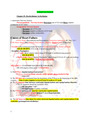
1. The P-Wave-depolarization (contraction)
+ P-Wave should be round, smooth, small, upright above isoelectric line
- Like a hill
2. The PR Interval is measured from the beginning of the P Wave to the beginning of the QRS complex. Time it takes impulse to travel from SA node to AV node
4. The ST Segment-time between ventricular depolarization and repolarization (diastole).
+ The ST Segment should be isoelectric (flat).
- ST elevation MI!!
- ST depression Cardiac ischemia
5. The T Wave represents the time for ventricular repolarization
+ T Wave should be upright [peaked T wave=hyperkalemia]
+ Inverted T wave=abnormality
6. The QT Interval-time taken for entire electrical depolarization and repolarization of the ventricles [prolonged=dysrhythmias]
Key Things to Remember:
+ Tiny box = 0.04 seconds
+ Big box = 5 tiny boxes = 0.2 seconds
+ PR Interval = 3-5 tiny boxes (0.12-0.2 seconds)
- PR interval > 5 boxes = 1st degree heart block
+ QRS = 1-3 tiny boxes (0.04-0.12 second)
- QRS > 3 boxes = some type of ventricular problem
+ ST depression = Ischemia
+ ST elevation = MI (worse than ischemia)
+ P wave = Atrial contraction (multiple p waves= a-fib stroke)
+ Increase HR, Decrease BP = Dehydration Can be sinus tachycardia
*** Signs and Symptoms of Decreased Cardiac Output ***
+ Decreased LOC (Confusion, Dizziness, Syncope, Restlessness, Agitation, Lethargy, Coma)
+ Muscle weakness, Angina, Decreased BP, SOB, Capillary Refill > 3 sec, hypoxia, decreased urine output, pale skin
*** Dysrhythmias Key Points to Remembers ***
+ Dysrhythmias are not treated unless the patients are symptomatic
+ Dysrhythmias can be categorized as either too fast, too slow, or too ugly
+ Atrial dysrhythmias are fast heart rates with narrow QRS complexes
+ Atrial dysrhythmias lead to strokes
+ Ventricular dysrhythmias have wide QRS complexes
+ When rhythms are slow, we want to increase HR
+ When rhythms are fast, we want to slow HR
+ When rhythm is pulseless ventricular tachycardia or ventricular fibrillation, the patient requires defibrillation in addition to CPR
+ Pulseless electrical activity is when there is a cardiac rhythm, but the patient
does not have a pulse
+ Asystole cannot be defibrillated
Changes Associated with Myocardial Ischemia
1. ST segment is depressed = Ischemia
- Normal ST line would be at isoelectric line level
2. T wave is inverted = MI (old)
Patients with ischemia may display one or both changes
Changes Associated with Injury
1. Physiologic Q wave- first negative deflection (wave) following the P wave. It is normally very short and narrow
2. Dramatic ST segment elevation = MI
Normal Sinus Rhythm
+ Sinus node fires 60-100 beats/minute
+ Follows normal conduction pattern
+ P wave upright and uniform & precedes QRS complex
+ PQ interval 3-5 boxes
+ QRS narrow 1-3 boxes and equal distance
- Distance same = Regular rhythm
+ T wave upright and uniform [Show Less]